Automatic pancreas detection in 3d CT scans
Multi-stage system of pancreas segmentation using CT scans.
Our developers have used Matlab code on multi-stage system of pancreas segmentation from CT scans. It allows to train a model on a large dataset of scans with corresponding label files acquired from a human expert, save produced predictive components, estimate their expected accuracy and apply the trained model for image segmentation.
Training algorithm was implemented according to the scheme described in “Deep convolutional networks for pancreas segmentation in CT imaging” paper by H. Roth, A. Farag, L. Lu, E. Turkbey and R. Summers.
Analysis includes preprocessing stage where 3-dimensional SLIC segmentation is performed in order to find image components/superpixels with homogeneous properties which will be later classified together.
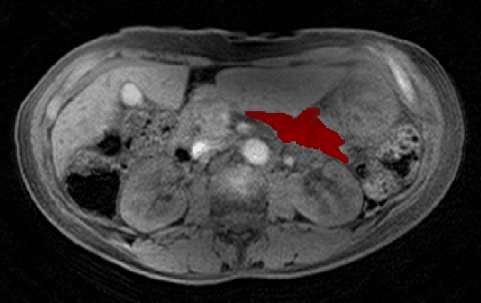
At the next stage, random forest classification model is trained to extract only that small region of interest that contains pancreas with high probability.
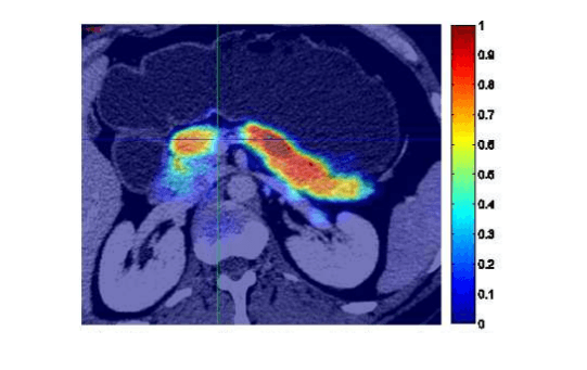
And finally, the deep convolutional neural network was trained to produce a refined probability of being pancreas for each superpixel.
Not all parameters of the system were explicitly provided in the paper, so the code was built to make it easy to modify them and test several combinations.
Code was optimized so that it can work efficiently on large image files and allow a fast update of a training set: only the parameters directly connected to the updated files would be recomputed.
Various visualizations and similarity measures were applied to confirm the correctness of the implementation on different data processing stages.
- Image processing
- Image segmentation
- Pattern recognition

Similar Projects
Mobile App for Connected Medical Hardware
iOS app for connected health devices with BLE integration and data reporting.
Multi-Channel AI Platform
A chatbot platform with NLP, built for flexible interactions, cross-channel deployment, and continuous product expansion.
AI-Powered Housekeeping
A mobile app that streamlines housekeeping workflows, tracks tasks in real time, and ensures consistent cleaning quality.